AKT/PI3K Signaling Pathway
AKT/PI3K is one of the most actively studied kinase pathways in basic research and drug development, as it plays an integral role in mediating signals for cell growth, survival, cell-cycle progression, differentiation, transcription, translation, and glucose metabolism (See figure 1). Recent advances in AKT signaling have focused on understanding cellular processes and identifying cellular substrates that are physiologically relevant in vivo. These efforts have uncovered important roles for AKT pathway regulation in cancer research, neuroscience, and disease prevention.
Also known as PKB, AKT is a serine/threonine kinase composed of an N-terminal regulatory domain, a hinge region that connects the regulatory domain to a kinase domain, and a C-terminal region required for the induction and maintenance of the kinase activity. Initially, AKT was discovered as a proto-oncogene. There are three highly homologous isoforms known as AKT1 or simple AKT, AKT2, and AKT3 with alternatively named PKB α, PKB β, and PKB γ, respectively. AKT1 plays an important role in cellular survival by inhibiting apoptotic processes and is implicated as a major factor in many types of cancer. AKT2 required to induce glucose transport is an important signaling molecule in the insulin signaling pathway. The role of AKT3 is less clear, though it appears to be predominantly expressed in the brain. Deregulations in the AKT-related pathway were observed in many human diseases, including cancer, cardiopathies, neurological disorders, and type-2 diabetes.
Thanks to the funding support from the National Cancer Institute (NCI Awards HHSN26100900070C and HHSN261201100087C), Rockland has developed over 100 AKT-related products. Recombinant AKT calibrator proteins have been produced as both AKT active (phosphorylated) and non-active (phosphatase-treated and phosphorylation site double mutant recombinant proteins). These reagents can be used for detecting status of proteins involved in AKT/PI3K pathway including AKT1, AKT2, and AKT3 isoforms and their phosphorylated stages.
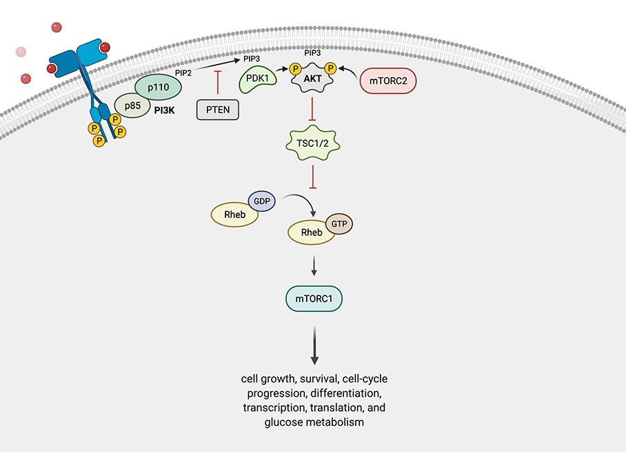
Figure 1: AKT/PI3K Pathway. Following generation of the second messenger PIP3 by PI3K, both phosphoinositide-dependent kinase-1 (PDK1) and AKT are recruited to the membrane. The proximity of these kinases allows PDK1 to phosphorylate AKT at residue T308 of the activation loop (T-loop). Subsequently, AKT is phosphorylated at residue S473 of the hydrophobic motif by the rapamycin-insensitive mTOR complex 2 (mTORC2). This phosphorylation is necessary to fully activate the kinase activity of AKT. AKT itself can phosphorylate several other substrates such as TSC2, a negative regulator of mTORC1 activity. (Adapted from Castel et al., 2014, created with BioRender.com)
Profiling AKT Protein Activation Status
Faulty activation of AKT underlies the pathophysiological properties of a variety of complex diseases, including type-2 diabetes and cancer. Constitutively activated, AKT promotes cellular survival as well as resistance to treatment with chemotherapy and radiation therapy. In fact, several research groups are developing compounds targeting AKT as anticancer drugs. Quantitation of the expression of AKT proteins and their activated status in response to the pathway inhibitors would have great potential application in monitoring therapeutic efficacy.
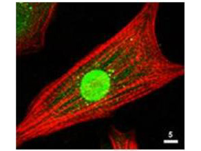
Figure 2: Localization of AKT (Green) by fluorescence microscopy using AKT Antibody
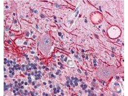
Figure 3: Localization of active AKT (Red) in human brain cerebellum tissue using AKT phospho T308 Antibody
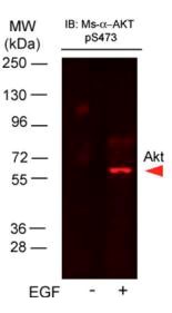
Figure 4: Western blot using A431 cells stimulated with EGF and AKT phospho S473 Antibody
Pharmacodynamic Assay for AKT Inhibitors
AKT/PI3K pathway has become a major focus in the research community thanks to its regulatory role in diverse cellular processes, including cell survival, cancer progression, and insulin metabolism. The AKT cascade is activated by a number of stimuli such as receptor tyrosine kinases, G-protein-coupled receptors, cytokine receptors integrins, and B and T cell receptors. In recent years, alongside the well-characterized AKT1 isoform, AKT2 and AKT3 have emerged as significant contributors to cancer with distinct non-overlapping functions. The AKT pathway is an attractive target for developing anti-cancer drugs as some experimental cancer drugs (AKT inhibitors e.g., Perifosine; mTOR inhibitors e.g., rapamycin; PI3K inhibitors e.g., LY294002) have been developed and used in clinical trials.
Isoforms of AKT are found to be distinct with regard to tissue expression, pathway activation, and inhibitor sensitivity. Rockland has created a panel of monoclonal antibodies to AKT isoforms. These anti-AKT antibodies are available to academic researchers as well as biopharma companies who wish to manufacture highly sensitive pharmacodynamic assays for measuring both total AKT, its isoforms, and levels of phosphorylated AKT in response to AKT inhibitors and modulators.
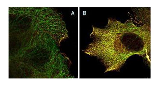
Figure 5: Immunofluorescence Microscopy of AKT phospho S473 Antibody using STED nanoscopy to evaluate AKT activation and migration. Panel A: serum starved, unstimulated A431 cells. Panel B: serum starved A431 cells with 15 minute EGF stimulation.
Quantitation of the expression of AKT proteins and their activated status as well as activation and expression status of other proteins involved in AKT pathway in response to the pathway inhibitors would have great potential application in monitoring therapeutic efficacy.
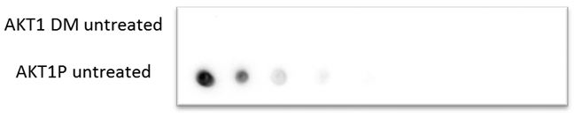
Figure 6: Dot blot illustrating reactivity of AKT phospho T308 Antibody
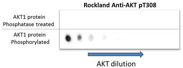
Figure 7: Dot blot demonstrating the phosphorylation status of AKT. Recombinant AKT was untreated or treated with phosphatase to remove the phosphorylation at the T308 and S473 sites. Only the phosphorylated AKT is detected by AKT phospho T308 Antibody.
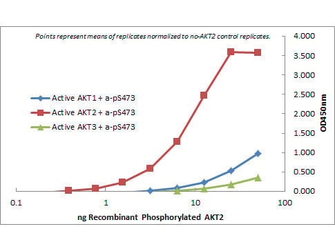
Figure 8: Sandwich ELISA assay to profiling active AKT2 Isoform
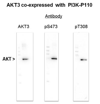
Figure 9: Recombinant AKT3 detected with AKT specific antibodies. AKT3 co-expressed with P110 Kinase leads to phosphorylation as demonstrated by reactivity with AKT phospho S473 Antibody and AKT phospho T308 Antibody.
AKT Antibodies for Cell Signaling
Activation of AKT results in phosphorylation of a wide range of proteins downstream of the AKT pathway, making AKT Western blotting a popular technique for monitoring this reaction. The phosphorylated substrates, present in various subcellular locations play an important role in directing different phenotypic outcomes. Faulty or aberrant activation of AKT underlies the pathophysiological properties of a variety of complex diseases, including type-2 diabetes, HIV, and cancer. When constitutively active, AKT is utilized frequently by cancer cells to circumvent therapeutic intervention, promoting cellular survival and resistance to chemo and radiation therapy. Our AKT antibodies and AKT/PI3K pathway reagents help monitor levels of AKT and its phosphorylation at position S473, T308, as well as AKT antibody isoforms AKT2 and AKT3.
AKT Antibodies
| Product | Clonality | Reactivity | Application |
| AKT phospho S473 Antibody | Monoclonal | Human, Mouse, Rat, Mokey | WB, IF, IHC, FC, ELISA |
| AKT phospho S473 Antibody | Polyclonal | Human, Mouse, Rat | WB, IF, IHC, Dot Blot, ELISA |
| AKT phospho T308 Antibody | Monoclonal | Human, Mouse | WB, IF, IHC, ELISA |
| AKT phospho T308 Antibody | Polyclonal | Human, Mouse, Rat | WB, IHC, Dot Blot, ELISA |
| AKT Antibody | Monoclonal | Human, Mouse, Chimpanzee, Rat | WB, ELISA |
| AKT Antibody | Polyclonal | Human, Mouse, Rat, Chicken | WB, IF, IHC |
| AKT1 Antibody | Monoclonal | Human, Mouse | WB, IP, FC, ELISA |
| AKT2 Antibody | Moncolonal | Human | WB, IHC, ELISA |
| AKT2 Antibody | Polyclonal | Human, Mouse | WB, ELISA |
| AKT3 Antibody | Monoclonal | Human | WB, IF, ELISA |
AKT/PI3K Pathway Antibodies
| Product | Clonality | Reactivity | Application |
| BAD Antibody | Polyclonal | Human, Mouse, Rat | WB, IF, IHC, ELISA |
| mTOR Antibody | Polyclonal | Human | WB, ELISA |
| PDK1 Antibody | Polyclonal | Human | WB, ELISA |
| PI 3 Kinase p85 alpha phospho Y607 Antibody | Polyclonal | Human | IHC, Dot Blot |
| PI3-kinase p110 delta Antibody | Polyclonal | Human, Mouse | WB |
| PTEN Antibody | Polyclonal | Human, Mouse | WB, IP, ELISA |
| Raptor Antibody | Polyclonal | Mouse | WB, IHC, ELISA |
| Rheb Antibody | Polyclonal | Human, Mouse, Rat | WB, IF, IHC, ELISA |
| TSC1 Antibody | Polyclonal | Human, Mouse, Rat | WB, IF, IHC, ELISA |
| TSC2 Antibody | Polyclonal | Human, Mouse | WB, ELISA |