You may also want to consider these related products.
TNF-α Mouse Recombinant Protein - 010-001-321
Anti-Human TNF p55 Receptor (sTNFRp55) (RABBIT) Antibody - 109-401-308
Anti-Mouse TNFα (RABBIT) Antibody - 110-401-321
Anti-Human TNF-α (RABBIT) Antibody - 209-401-306
Primary Antibodies  >  Stem Cell Antibodies
 

TNF alpha Antibody

Rabbit Polyclonal
Code

210-401-321
Size

1 mg
Price

$294.00
Availability

Ships within 2 weeks.
Order Offline:
Phone: 800.656.7625
E-mail: orders@rockland-inc.com
Fax: 610.367.7825
Bulk Orders:
Call or E-mail Us For Information
Fill out our Contact Form
Shipping & Handling Fees Apply.

Shipping:
$85.00 to United States
Handling:
$45.00
Synonyms: APC1 antibody, Cachectin antibody, DIF antibody, Differentiation inducing factor antibody, Macrophage cytotoxic factor antibody, MCF antibody, Necrosin antibody, TNF-alpha, Tumor necrosis factor ligand superfamily member 2, TNF-a, TNFA

TNF alpha Antibody Properties

Description
Anti-Mouse TNF-a (RABBIT) Antibody - 210-401-321
Host
Rabbit
Target Species
Mouse
Known Cross Reactivity
mouse
Clonality
Polyclonal
Application
ELISA : 1:1,000 - 1:5,000
Western Blot : 1:500 - 1:2,000
Other Dilution: User Optimized
Physical State
Liquid (sterile filtered)
Shipping condition
Dry Ice
Format
IgG
Label
Concentration
1.1 mg/mL by UV absorbance at 280 nm
Buffer
0.02 M Potassium Phosphate, 0.15 M Sodium Chloride, pH 7.2
Preservative
0.01% (w/v) Sodium Azide
Stabilizer
None
GeneName
TNFA
Download CofA
Lot#  

TNF alpha Antibody Description

Background
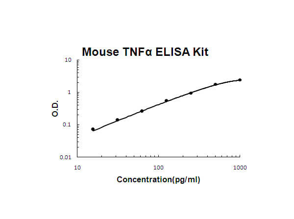
TNF alpha Antibody detects TNF-a protein. TNF-a is a cytokine that binds to TNFRSF1A/TNFR1 and TNFRSF1B/TNFBR. It is mainly secreted by macrophages and can induce cell death of certain tumor cell lines. It is a potent pyrogen causing fever by direct action or by stimulation of interleukin-1 secretion and is implicated in the induction of cachexia, Under certain conditions it can stimulate cell proliferation and induce cell differentiation. Anti-TNF-alpha Antibody is ideal for investigators involved in Cell Signaling, Immunology and Signal Transduction research.
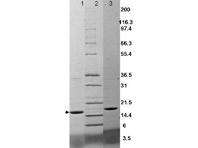
Immunogen
This antibody was produced from whole rabbit serum prepared by repeated immunizations with recombinant mouse TNFa produced in E.coli.
Immunogen Type
Recombinant Protein
Storage Condition
Store vial at -20° C prior to opening. Aliquot contents and freeze at -20° C or below for extended storage. Avoid cycles of freezing and thawing. Centrifuge product if not completely clear after standing at room temperature. This product is stable for several weeks at 4° C as an undiluted liquid. Dilute only prior to immediate use.
Application Note
This antibody against Mouse TNFa has been tested for use in neutralizations, ELISA, immunohistochemistry, radioimmunoassay, immunoprecipitation, immunohistochemistry and immunoblotting.  It recognizes the 17,000 MW TNFa.  Reactivity in other immunoassays is unknown. This antibody will recognize the cell-bound precursor of TNFa as a 26,000 protein in immunoblots, particularly in denatured samples. This antibody is also useful for neutralization of mouse activity in bioassays. It does not neutralize the biological activity of lymphotoxin. For neutralization, it is recommended to incubate the sample with a 1:200 dilution of the antibody for at least 4 hours before being tested. A control of similarly diluted normal rabbit IgG is recommended.
Purity/Specifity
TNF alpha Antibody has been heated to 56°C for 30 minutes.  The antibody is directed against mature 17,000 MW mouse TNFa and is useful in determining its presence in various assays. The antibody does not recognize mouse TNFß (lymphotoxin).
Disclaimer Note-General
This product is for research use only and is not intended for therapeutic or diagnostic applications. Please contact a technical service representative for more information. All products of animal origin manufactured by Rockland Immunochemicals are derived from starting materials of North American origin. Collection was performed in United States Department of Agriculture (USDA) inspected facilities and all materials have been inspected and certified to be free of disease and suitable for exportation. All properties listed are typical characteristics and are not specifications. All suggestions and data are offered in good faith but without guarantee as conditions and methods of use of our products are beyond our control. All claims must be made within 30 days following the date of delivery. The prospective user must determine the suitability of our materials before adopting them on a commercial scale. Suggested uses of our products are not recommendations to use our products in violation of any patent or as a license under any patent of Rockland Immunochemicals, Inc. If you require a commercial license to use this material and do not have one, then return this material, unopened to: Rockland Inc., P.O. BOX 326, Gilbertsville, Pennsylvania, USA.
General Reference
Specific Reference
There are no reviews for this product. Log in to submit your own review.

AMCA Conjugates
Antigen Retrieval Methods
Azide Removal by Dialysis
Beta Galactosidase Conjugates
Biotin Binding Assay (HABA)
Biotin Conjugates
Cell Lysates Protocol
CyDye™ Conjugates
DyLight™ Antibody Conjugate Properties
DyLight™ Antibody Spectra
ELISA
ELISA to Quantify Collagen
Extended Conjugate Information Sheet
Fab Fragment Monovalent Antibodies
FACS / Flow Cytometry Analysis
Fluorescein Conjugates
Gel Shift Assay
General Antibody Information
Histone Immunoblotting
Immunoblotting
Immunocytochemistry
Immunofluorescence Microscopy
Immunofluorescence Microscopy - Assay Formats
Immunofluorescence Microscopy - ATTO-TEC
Immunofluorescence Microscopy - CyDye®
Immunofluorescence Microscopy - DyLight™
Immunofluorescence Microscopy - Fluorescent Labels
Immunofluorescence Microscopy - IRDye®
Immunofluorescence Microscopy - long
Immunofluorescence Microscopy - Microscope Types
Immunofluorescence Microscopy - Other Conjugates
Immunofluorescence Microscopy Protocol
Immunohistochemistry
Immunoprecipitation
Nuclear/Cytoplasmic Extract Isolation
Peroxidase & Alkaline Phosphatase
Phosphorylated State Proteins
Phosphorylated State Proteins - AKTs
Phosphorylated State Proteins - Long
Phosphorylated State Proteins - Pathways
Phosphorylated State Proteins - Phospho Specific
Phosphorylated State Proteins - Tailored Phosphorylated Target
Phosphorylated State Proteins - Western Blot
Phycoerythrin Conjugates
Protein A / Protein G Binding Reactivities
Recommended Dilutions
Rhodamine & Texas Red™
SDS-PAGE
Staining Paraffin Sections by PAP Procedure
USDA & Material Safety Notice
Western Blot - Buffers
Western Blot - Long
Western Blot - Preassembled Kits
Western Blot - Primary Antibody Reagents
Western Blot - Secondary Antibody Reagents
Western Blot - Substrates
Western Blot - Types of Blot Formats
Western Blot (Protein Immunoblot)
Product Type
Proteins and Peptides;
Reacts With
Mouse
Catalog Number
010-001-321

Product Type
Primary Antibodies;
Applications
ELISA, WB
Reacts With
human, primate
Catalog Number
109-401-308

Product Type
Primary Antibodies;
Applications
ELISA, WB
Reacts With
mouse
Catalog Number
110-401-321

Product Type
Primary Antibodies;
Applications
ELISA, WB, IHC
Reacts With
human
Catalog Number
209-401-306

Product Label


Description
Unconjugated
Color
Conjugation Reference
Molecular Weight
Excitation Wavelength
Conjugation Chemistry