


HEF1 Antibody
Mouse Monoclonal 2G9 IgG1 kappa
$50.00 to US & $70.00 to Canada for most products. Final costs are calculated at checkout.
Background
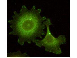
HEF1, also known as Enhancer of filamentation 1, CRK-associated substrate-related protein, CAS-L, CasL, p105 and Neural precursor cell expressed developmentally down-regulated 9 is the product of the NEDD9 (CASGL) gene. HEF1 functions as a docking protein that plays a central coordinating role for tyrosine-kinase-based signaling related to cell adhesion. HEF1 may also function in transmitting growth control signals between focal adhesions at the cell periphery and the mitotic spindle in response to adhesion or growth factor signals initiating cell proliferation. HEF1 may also play an important role in integrin beta-1 or B cell antigen receptor (BCR) mediated signaling in B- and T-cells. Integrin beta-1 stimulation leads to recruitment of various proteins including CRK, NCK and SHPTP2 to the tyrosine phosphorylated form. HEF1 forms a homodimer and can heterodimerize with HLH proteins ID2, E12, E47 and also with p130cas. HEF1 also forms complexes in vivo with related adhesion focal tyrosine kinase (RAFTK), adapter protein CRKL and LYN kinase and also interacts with MICAL and TXNL4/DIM1. This protein localizes to both the cell nucleus and the cell periphery and is differently localized in fibroblasts and epithelial cells. In fibroblasts, it is predominantly nuclear and in some cells is present in the Golgi apparatus. In epithelial cells, it is localized predominantly in the cell periphery with particular concentration in lamellipodia, but it is also found in the nucleus. HEF1 is widely expressed although higher levels are detected in kidney, lung, and placental tissue. HEF1 is also detected in T-cells, B-cells and diverse cell lines. HEF1 is activated upon induction of cell growth. Cell cycle-regulated processing produces four isoforms: p115, p105, p65, and p55. Isoform p115 arises from p105 phosphorylation and appears later in the cell cycle. Isoform p55 arises from p105 as a result of cleavage at a caspase cleavage-related site and it appears specifically at mitosis. The p65 isoform is poorly detected. Isoforms p105 and p115 are predominantly cytoplasmic and associate with focal adhesions while p55 associates with the mitotic spindle.
Product Details
Target Details
Application Details
Formulation
Shipping & Handling
This product is for research use only and is not intended for therapeutic or diagnostic applications. Please contact a technical service representative for more information. All products of animal origin manufactured by Rockland Immunochemicals are derived from starting materials of North American origin. Collection was performed in United States Department of Agriculture (USDA) inspected facilities and all materials have been inspected and certified to be free of disease and suitable for exportation. All properties listed are typical characteristics and are not specifications. All suggestions and data are offered in good faith but without guarantee as conditions and methods of use of our products are beyond our control. All claims must be made within 30 days following the date of delivery. The prospective user must determine the suitability of our materials before adopting them on a commercial scale. Suggested uses of our products are not recommendations to use our products in violation of any patent or as a license under any patent of Rockland Immunochemicals, Inc. If you require a commercial license to use this material and do not have one, then return this material, unopened to: Rockland Inc., P.O. BOX 5199, Limerick, Pennsylvania, USA.