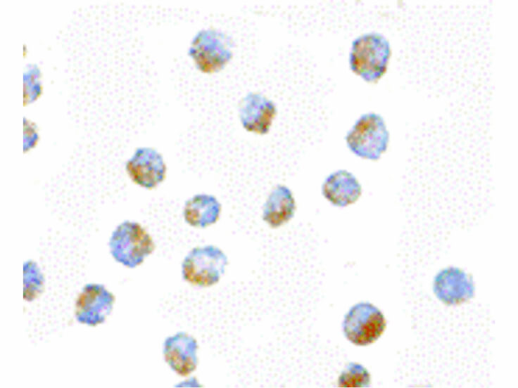
You may also want to consider these related products.
Anti-Cyclin E (RABBIT) Antibody - 100-401-156
Anti-cdk2 (RABBIT) Antibody - 100-401-161
Anti-BUBR1 Kinase (MOUSE) Monoclonal Antibody - 200-301-902
Anti-MOUSE IgG (H&L) (RABBIT) Antibody Peroxidase Conjugated - 610-4302
Primary Antibodies  >  Cell Cycle Protein Antibodies
 

BUB1 Antibody

Mouse Monoclonal 14H5 IgG1
Code

200-301-900
Size

100 µg
Price

$294.00
Availability

Ships next business day.
Order Offline:
Phone: 800.656.7625
E-mail: orders@rockland-inc.com
Fax: 610.367.7825
Bulk Orders:
Call or E-mail Us For Information
Fill out our Contact Form
Shipping & Handling Fees Apply.

Shipping:
$85.00 to United States
Handling:
$45.00
Synonyms: Budding uninhibited by benzimidazoles 1 homolog antibody, hBUB1 antibody, Mitotic checkpoint serine/threonine protein kinase BUB1 antibody

BUB1 Antibody Properties

Description
Anti-BUB1 (MOUSE) Monoclonal Antibody - 200-301-900
Host
Mouse
Target Species
Human
Known Cross Reactivity
human
Clonality
Monoclonal 14H5 IgG1
Application
ELISA : 1:5,000 - 1:20,000
IF Microscopy : 1:500 - 1:2,000
Western Blot : 1:500 - 1:2,000
ImmunoPrecipitation: 1:200
Other Dilution: User Optimized
Physical State
Liquid (sterile filtered)
Shipping condition
Dry Ice
Format
IgG
Label
Concentration
1.0 mg/mL by UV absorbance at 280 nm
Buffer
0.02 M Potassium Phosphate, 0.15 M Sodium Chloride, pH 7.2
Preservative
0.01% (w/v) Sodium Azide
Stabilizer
None
GeneName
BUB1
Download CofA
Lot#  

BUB1 Antibody Description

Background
BUB1 (also called Mitotic checkpoint serine/threonine-protein kinase BUB1and BUB1A) is a kinase involved in spindle checkpoint function. The kinase functions in part by phosphorylating BUB3, a member of the miotic checkpoint complex, and activating the spindle checkpoint.  Mutations in this gene have been associated with aneuploidy and several forms of cancer.   BUB1 is autophosphorylated when cells enter mitosis.  This protein is localized to the nucleus in interphase cells. Kinetochore localization is required for normal mitotic timing and checkpoint response to spindle damage. BUB1 is highly expressed in testis and thymus, less in colon, spleen, lung and small intestine.  Expression is associated with cells/tissues with a high mitotic index.
Immunogen
This protein A purified monoclonal antibody was produced by repeated immunizations with a recombinant protein corresponding to amino acid residues 281-419 of human BUB1 protein.
Immunogen Type
Recombinant Protein
Storage Condition
Store vial at -20° C prior to opening.  Aliquot contents and freeze at -20° C or below for extended storage.  Avoid cycles of freezing and thawing.  Centrifuge product if not completely clear after standing at room temperature.  This product is stable for several weeks at 4° C as an undiluted liquid.  Dilute only prior to immediate use.
Application Note
This protein A purified antibody has been tested for use in immunoprecipitation, immunofluorescence staining and western blot and is capable of detecting endogenous protein.  Specific conditions for reactivity should be optimized by the end user.  Expect a predominant band at ~ 160 kDa corresponding to full-length protein by western blotting in the appropriate cell lysate or extract.  The use of HeLa whole cell lysates prepared using a RIPA buffer is recommended as a positive control.  For IF microscopy use cells grown on cover slips fixed with 3.5% paraformaldehyde in PBS at pH 6.8 OR 100% methanol at -20° C.  Permeabilize fixed cells with 0.5% Triton X-100.
Purity/Specifity
This Protein A purified antibody is directed against human BUB1 protein. The product was purified from tissue culture supernatant by chromatography.  This antibody reacts with BUB1 from human cells.  No reactivity is seen for homologues from Xenopus or Kangaroo Rat.    Reactivity against homologues from other sources is not known.
Disclaimer Note-General
This product is for research use only and is not intended for therapeutic or diagnostic applications. Please contact a technical service representative for more information. All products of animal origin manufactured by Rockland Immunochemicals are derived from starting materials of North American origin. Collection was performed in United States Department of Agriculture (USDA) inspected facilities and all materials have been inspected and certified to be free of disease and suitable for exportation. All properties listed are typical characteristics and are not specifications. All suggestions and data are offered in good faith but without guarantee as conditions and methods of use of our products are beyond our control. All claims must be made within 30 days following the date of delivery. The prospective user must determine the suitability of our materials before adopting them on a commercial scale. Suggested uses of our products are not recommendations to use our products in violation of any patent or as a license under any patent of Rockland Immunochemicals, Inc. If you require a commercial license to use this material and do not have one, then return this material, unopened to: Rockland Inc., P.O. BOX 326, Gilbertsville, Pennsylvania, USA.
General Reference
Specific Reference
There are no reviews for this product. Log in to submit your own review.

AMCA Conjugates
Antigen Retrieval Methods
Azide Removal by Dialysis
Beta Galactosidase Conjugates
Biotin Binding Assay (HABA)
Biotin Conjugates
Cell Lysates Protocol
CyDye™ Conjugates
DyLight™ Antibody Conjugate Properties
DyLight™ Antibody Spectra
ELISA
ELISA to Quantify Collagen
Extended Conjugate Information Sheet
Fab Fragment Monovalent Antibodies
FACS / Flow Cytometry Analysis
Fluorescein Conjugates
Gel Shift Assay
General Antibody Information
Histone Immunoblotting
Immunoblotting
Immunocytochemistry
Immunofluorescence Microscopy
Immunofluorescence Microscopy - Assay Formats
Immunofluorescence Microscopy - ATTO-TEC
Immunofluorescence Microscopy - CyDye®
Immunofluorescence Microscopy - DyLight™
Immunofluorescence Microscopy - Fluorescent Labels
Immunofluorescence Microscopy - IRDye®
Immunofluorescence Microscopy - long
Immunofluorescence Microscopy - Microscope Types
Immunofluorescence Microscopy - Other Conjugates
Immunofluorescence Microscopy Protocol
Immunohistochemistry
Immunoprecipitation
Nuclear/Cytoplasmic Extract Isolation
Peroxidase & Alkaline Phosphatase
Phosphorylated State Proteins
Phosphorylated State Proteins - AKTs
Phosphorylated State Proteins - Long
Phosphorylated State Proteins - Pathways
Phosphorylated State Proteins - Phospho Specific
Phosphorylated State Proteins - Tailored Phosphorylated Target
Phosphorylated State Proteins - Western Blot
Phycoerythrin Conjugates
Protein A / Protein G Binding Reactivities
Recommended Dilutions
Rhodamine & Texas Red™
SDS-PAGE
Staining Paraffin Sections by PAP Procedure
USDA & Material Safety Notice
Western Blot - Buffers
Western Blot - Long
Western Blot - Preassembled Kits
Western Blot - Primary Antibody Reagents
Western Blot - Secondary Antibody Reagents
Western Blot - Substrates
Western Blot - Types of Blot Formats
Western Blot (Protein Immunoblot)
Product Type
Primary Antibodies;
Applications
ELISA, WB
Reacts With
Human
Catalog Number
100-401-156

Product Type
Primary Antibodies;
Applications
ELISA, WB, IHC
Reacts With
human, rat and mouse
Catalog Number
100-401-161

Product Type
Primary Antibodies;
Applications
ELISA, WB, IF
Reacts With
human
Catalog Number
200-301-902

Product Type
Secondary Antibodies;
Applications
ELISA, WB, IHC
Catalog Number
610-4302

Product Label


Description
Unconjugated
Color
Conjugation Reference
Molecular Weight
Excitation Wavelength
Conjugation Chemistry